
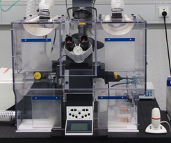
Leica SP5-STED confocal Laser scanning microscope
The Leica SP5 confocal laser scanning microscope is used for imaging of fluorescently labeled biological systems. Exploring morphology and phase separation of labeled polymers is another field of application. As a tandem system the confocal microscope can also image reflected light, extending the operation to non-fluorescent samples. In standard fluorescence confocal microscopy, the resolution is limited by the finite beam size of the excitation laser in the focus. Actually, the minimum diameter of the focus point is limited by the Abbé Limit to approximately half the laser-wavelength. In addition to the primary exitation beam, the STED module imposes a second, high intensity beam with a Donut-pattern. Its wavelength is chosen to match the long wavelength tail of the fluorophore emission. This leads to stimulated emission depletion of the fluorophores in the outer region of the spot that is illuminated by the focused excitation beam. The detector only registers spontaneous fluorescence from the short wavelength spectral emission, thus limiting the spatial origin of the signal to regions below the diffraction barrier. With increasing intensity of the Donut beam, the size of the signal rendering spot is decreasing. Therefore resolution scales with STED intensity and high intensity of the STED beam is essential.

Specifications
| Microscope design: | inverse |
| Objective lenses: | HC PL APO CS 10x/0.4 dry HCX PL APO CS 40x/0.85 dry HCX PL APO CS 40x/1.25-0.75 oil HCX PL APO CS 63x/1.4-0.6 oil HCX PL APO CS 63x/1.2 water HCX PL APO 63x/1.3 gylcerine 37°C corr. HCX PL APO CS 100x/1.4 oil |
| Excitation lasers: | Argon: 458nm,476nm,488nm,496nm,514nm DPSS: 561nm HeNe: 594nm,633nm |
| Detectors: | 4xPMT, 1xHyD, 2xAPD, 1xTransmission |
| Detection: | Spectrometer prism, (slit >=5nm), 400-780nm |
| Scan Range X/Y | 60-40mm |
| Scan line frequency | 1-16000Hz |
| Detectors: |
incubation chamber for live cell imaging (37°C, 5% CO2) |