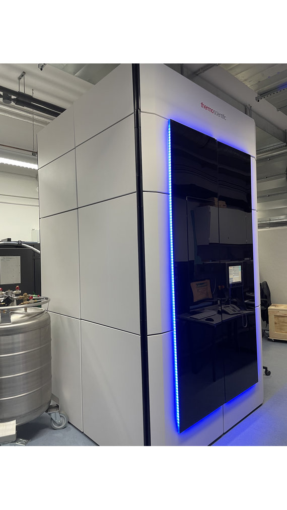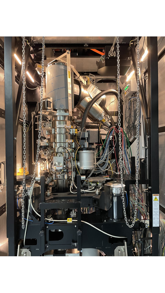
Krios G4 Cryo-TEM
An electron microscope uses electrons to produce a magnified image of a sample. In doing so, it achieves an extremely high resolution, which makes it possible to image even the atoms in the examined sample. However, for this to work, the sample under investigation must meet certain requirements: For example, the sample must not change in the vacuum of the microscope. Water in particular, which is essential for the structural integrity of biological molecules, would evaporate immediately in the vacuum of the electron microscope, leading to drastic changes in the structure under investigation.
A new electron microscope has now been installed at the Max Planck Institute for Polymer Research in which the sample is shock-frozen at -190 °C - a so-called "cryo" electron microscope (from the Greek "kryos": cold). The result is that the water can no longer evaporate, allowing biological systems to be studied in their "natural" environment.
In addition, the microscope installed at MPI-P was combined with an energy spectrometer, making it unique in the world. The spectrometer measures the energy of the electrons and thus allows conclusions to be drawn about the atoms present in the sample. This enables the researchers at the MPI-P to measure the chemical composition of the molecules under investigation with almost atomic resolution.

