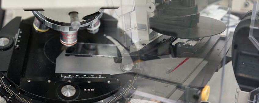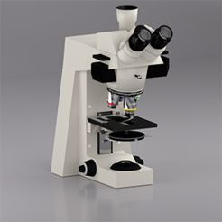
The optical microscope is a device which uses visible light as a probe in order to magnify the image of small objects. In principle, it works like any other transmission microscope. A light source emits the probe, usually white light. This light is then guided through the condenser system, which bundles the illumination to achieve a parallel light beam that is passed through the specimen. Accordingly, the specimen has to be transparent for the respective wave length. After the light passed the specimen, it enters the objective lens system, which provides for the first magnification. The quality of the objective lens is crucial for the following imaging system, because it defines the magnification and the attainable resolution of the microscope. Following the objective system, the light is guided to the eyepiece of the microscope, where the final magnified image of the sample is formed.

It is still doubtful, to whom the invention of the light microscope has to be ascribed. Clear is, that the first reports on two lens magnification systems dates back to the beginning of the 17th century. Since that time the optical microscopy was steadily developed and improved. Over the last four centuries the evolution produced astounding different microscopes with very specific areas of application, ranging from surgical to ultra microscopes, to name a few. Furthermore, the number of contrast techniques – that´s the main physical principle on which the image formation is based on – increased significantly. Some of the contrasts used in optical microscopy are phase, polarization, differential and fluorescence contrast, just to name a few. With the development of compact and reliable laser sources a rather new and powerful new class of optical microscopes evolved, the confocal laser scanning microscopes.
The maximum achievable resolution, however, is universal for all light based microscopes. It arises from the wave nature of the used light probe. Due to diffraction effects at the (necessary) apertures and at the rim of the lenses the maximum achievable resolution d is given by the Abbé Limit. As a rule over the thumb the maximum resolution is approximately half the wavelength of the used light, d ≈ 250 nm.
For the last four centuries, the Abbé Limit was axiomatic and breaking the resolution limit of optical microscopy seemed to be impossible. However, smart approaches in either technical as well as in labeling methods led to resolution limit breaking optical microscopy systems like STED, STORM and PALM. These developments were very important for imaging e.g. proteins in biological systems, that it was awarded the chemistry nobel price 2014.
At the EM-group we are operating three light optical microscopes, each for a very different application. The Zeiss Axiophot is a dedicated material science oriented microscope. Imaging nanoparticles down to a size of approximately 10 nm is featured by an ultramicroscope with a cyto-viva darkfield condenser system. Finally, we operate a confocal laser scanning microscope (Leica SP5) that is upgraded with a commercial extension for superresolution. It is mainly used as a standard confocal microscope for imaging fluorescently labeled biological systems. Exploring morphology and phase separation of labeled polymers is another field of application. As a tandem system the confocal microscope can also image reflected light, extending the operation to non-fluorescent samples.
